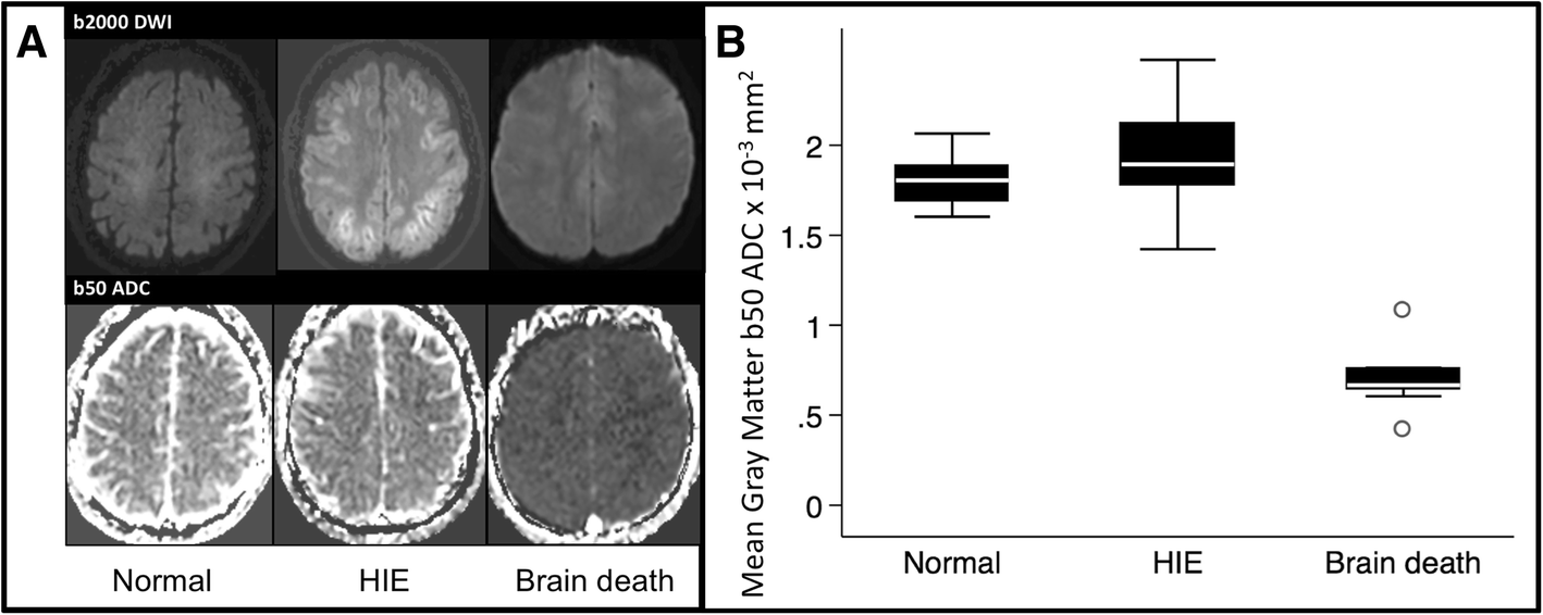
Fig. 1

b50 apparent diffusion coefficient (ADC) values in normal, hypoxic ischemic encephalopathy (HIE), and brain death populations. A panel of representative images (a) demonstrates high b-value DWI trace images (top) and b50 ADC maps (bottom). In brain death patients, there was diffuse hypointensity on ADC maps compared with normal and HIE subjects. Pooled quantified ADC data (b) in box-and-whisker format demonstrate markedly lower mean ADC values in brain death subject gray matter compared with normal and HIE subjects, p < 0.001
